×
General Ultrasound Imaging
-
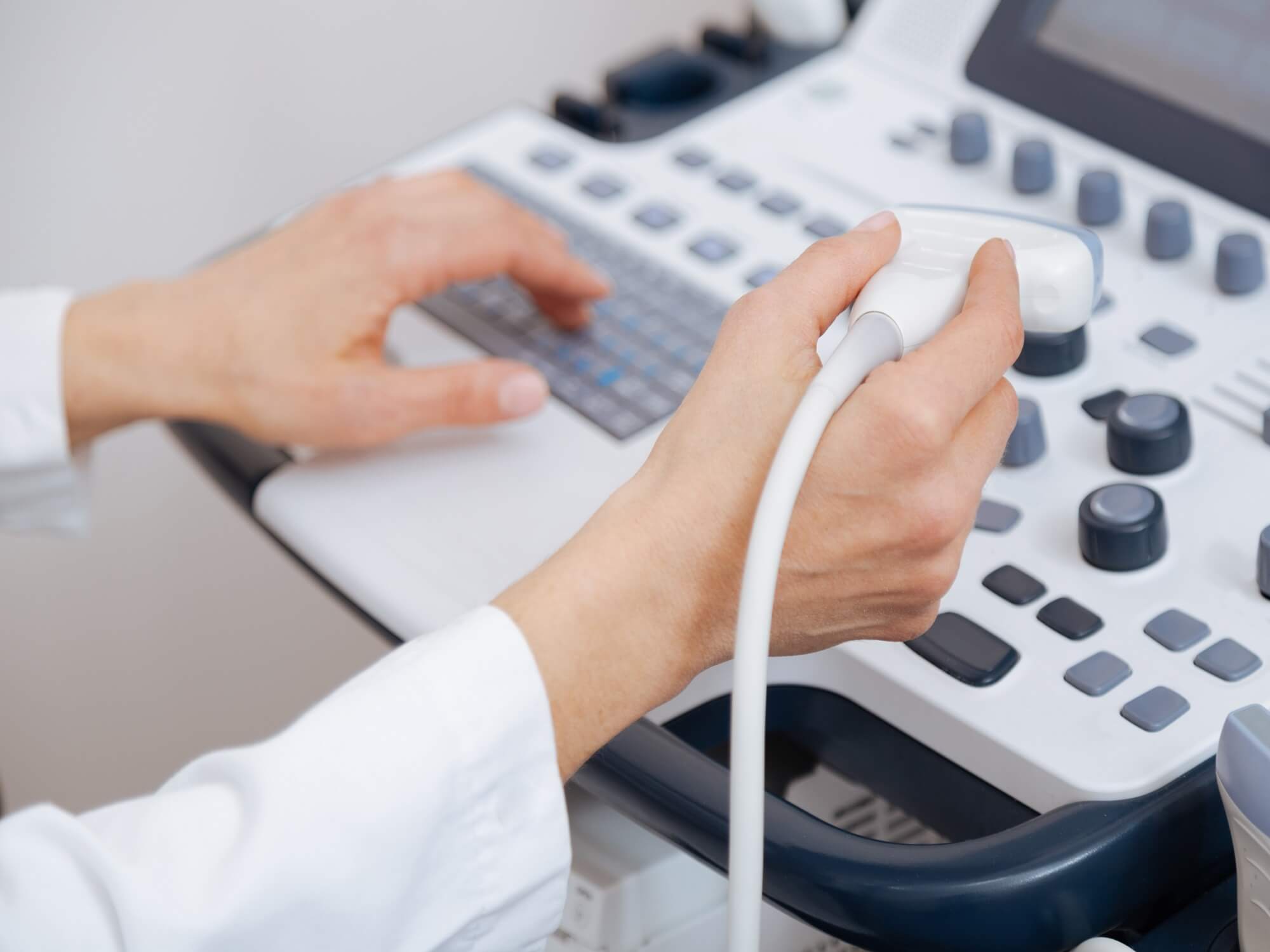
Ultrasound imaging, also called ultrasound scanning or sonography, involves the use of a small transducer (probe) and ultrasound gel to expose the body to high -frequency sound waves. Ultrasound is safe and painless, and produces pictures of the inside of the body using sound waves. Ultrasound examinations do not use Ionizing radiation (as used in x-rays). Because ultrasound images are captured in real-time, they can show the structure and movement of the body's internal organs, As well as blood flowing through blood vessels.
Conventional ultrasound displays the images in thin, flat sections of the body. Advancements in ultrasound technology include three-dimensional (3-D) ultrasound that formats the sound wave data into 3-D images. Four-dimensional (4-D) ultrasound is 3-D ultrasound in motion.
Doppler ultrasound is a special ultrasound technique that evaluates blood flow through a blood vessel, including the body's major arteries and veins in the abdomen, arms, legs and neck.
-
Why should I do it ?
-
Ultrasound is a useful way of examining many of the body's internal organs, including but not limited to the:
- Heart and blood vessels, including the abdominal aorta and its major branches
- Liver
- Gallbladder
- Spleen
- Pancreas
- Kidneys
- Bladder
- Uterus, ovaries, and unborn child (fetus) in pregnant patients
- Eyes
- Thyroid and parathyroid glands
- Scrotum (testicles(
- Brain in infants
- Hips in infants
-
Ultrasound is also used to:
- Guide procedures such as needle biopsies, in which needles are used to extract sample cells from an abnormal area for laboratory testing.
- Image the breasts and to guide biopsy of breast cancer (see the Ultrasound-Guided Breast Biopsy page.
- Diagnose a variety of heart conditions and to assess damage after a heart attack or diagnose for valvular heart disease.
-
Doppler ultrasound images can help the physician to see and evaluate:
- Blockages to blood flow (such as clots.
- Narrowing of vessels.
- Tumors and congenital vascular malformation.
-
Any preparations needed?
- You should wear comfortable, loose-fitting clothing.
- You may need to remove all clothing and jewelry in the area to be examined.
- Other preparation depends on the type of examination you will have. For some scans your doctor may instruct you not to eat or drink for as many as 12 hours before your appointment. For others you may be asked to drink up to six glasses of water two hours prior to your exam and avoid urinating so that your Bladder is full when the scan begins.
- In case of children, ultrasound examinations are very sensitive to motion, and an active or crying child will slow the examination process. To ensure a smooth experience, it would be beneficial to explain the procedure to the child prior to the exam.
-
Ultrasound is a useful way of examining many of the body's internal organs, including but not limited to the: